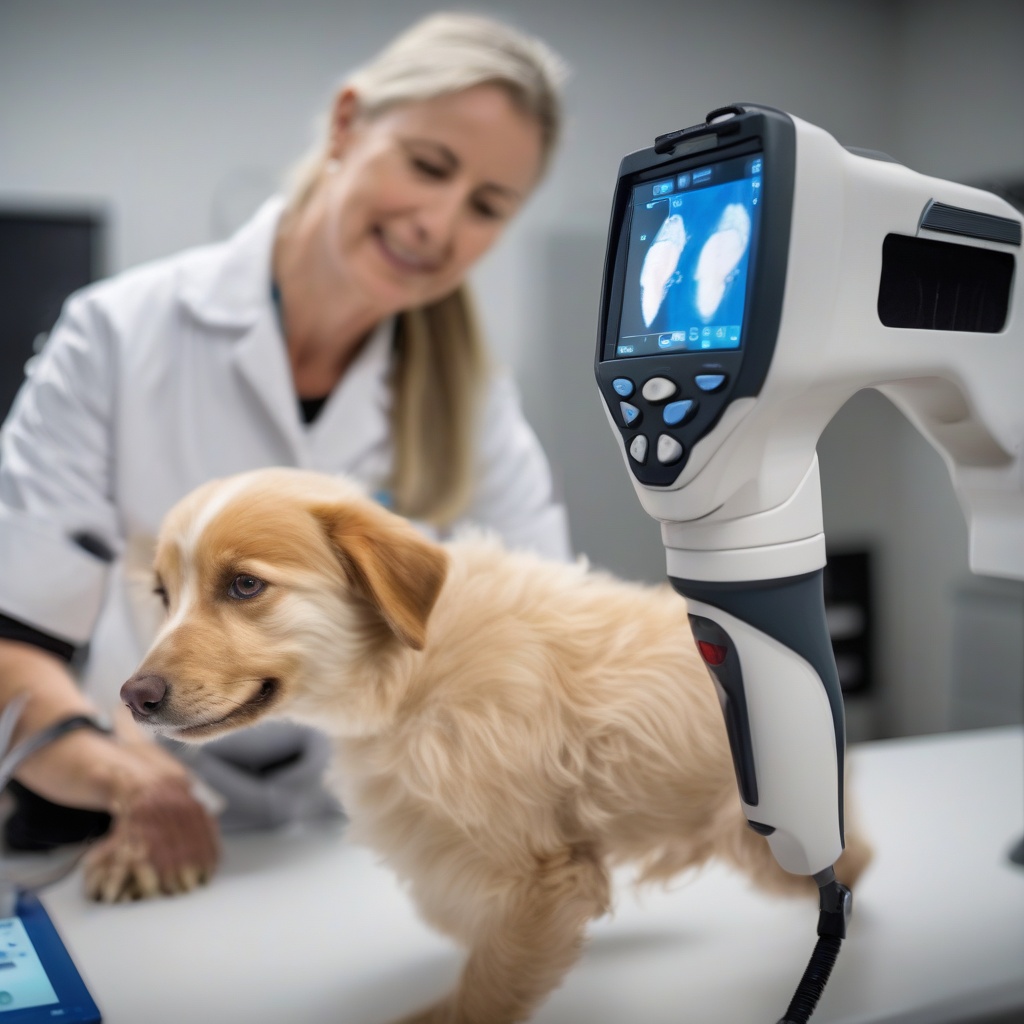
Expert Insights: Diagnosing Mast Cell Tumors with Advanced Thermal Imaging Techniques Revealed
Diagnosing mast cell tumors in pets can be a challenging and complex process. However, with the advancement of thermal imaging techniques, veterinarians can now make high-confidence diagnoses. In this blog post, we will explore the latest insights from experts in the field and discuss what to expect from the upcoming Fetch National Harbor event.
The Challenges of Diagnosing Mast Cell Tumors
Mast cell tumors are one of the most common types of skin tumors in dogs and cats. However, diagnosing them can be tricky, as they can resemble other skin conditions. Traditional diagnostic methods, such as fine-needle aspiration and histopathology, can be invasive and may not always provide a definitive diagnosis.
Advanced Thermal Imaging Techniques
Thermal imaging, also known as thermography, is a non-invasive imaging technique that uses infrared radiation to detect temperature differences in the body. In the context of mast cell tumors, thermal imaging can help identify abnormal heat patterns that may indicate the presence of a tumor.
Recent advances in thermal imaging technology have improved its sensitivity and specificity, making it a valuable tool for veterinarians. By using advanced thermal imaging techniques, veterinarians can:
- Detect mast cell tumors at an early stage, when they are more treatable
- Differentiate between mast cell tumors and other skin conditions
- Monitor the effectiveness of treatment and detect any potential recurrence
Expert Insights from Fetch National Harbor
The upcoming Fetch National Harbor event, taking place on September 17th, 2025, will feature a session with Dr. Zachary Wright, DVM, DACVIM, Oncology. Dr. Wright will share his expertise on using advanced thermal imaging techniques for diagnosing mast cell tumors.
In a recent interview, Dr. Wright discussed the benefits of thermal imaging in veterinary oncology:
“Thermal imaging is a game-changer for diagnosing mast cell tumors. It’s non-invasive, painless, and can provide valuable information about the tumor’s biology. By using thermal imaging, we can make more accurate diagnoses and develop more effective treatment plans.”
What to Expect from the Fetch National Harbor Event
The Fetch National Harbor event will bring together veterinarians and experts in the field to share knowledge and discuss the latest advancements in veterinary medicine. During the session with Dr. Wright, attendees can expect to learn:
- The principles of thermal imaging and its applications in veterinary oncology
- How to use thermal imaging to diagnose mast cell tumors and monitor treatment response
- Case studies and examples of successful thermal imaging applications in veterinary practice
For more information on the event and to learn more about Dr. Wright’s session, check out this link.
Conclusion
Advanced thermal imaging techniques are revolutionizing the diagnosis of mast cell tumors in pets. By attending the Fetch National Harbor event and learning from experts like Dr. Zachary Wright, veterinarians can stay up-to-date on the latest advancements in thermal imaging and improve their diagnostic confidence. Don’t miss this opportunity to enhance your knowledge and skills in veterinary oncology.


